Gadolinium »
PDB 6pou-7tso »
7lji »
Gadolinium in PDB 7lji: Structure of Poly(Aspartic Acid) Hydrolase PAHZ2 with Gd+3 Bound
Protein crystallography data
The structure of Structure of Poly(Aspartic Acid) Hydrolase PAHZ2 with Gd+3 Bound, PDB code: 7lji
was solved by
C.A.Brambley,
T.J.Yared,
M.Gonzalez,
A.L.Jansch,
J.R.Wallen,
M.H.Weiland,
J.M.Miller,
with X-Ray Crystallography technique. A brief refinement statistics is given in the table below:
| Resolution Low / High (Å) | 98.57 / 1.85 |
| Space group | P 21 21 21 |
| Cell size a, b, c (Å), α, β, γ (°) | 48.67, 147.87, 197.14, 90, 90, 90 |
| R / Rfree (%) | 14.5 / 16.2 |
Gadolinium Binding Sites:
The binding sites of Gadolinium atom in the Structure of Poly(Aspartic Acid) Hydrolase PAHZ2 with Gd+3 Bound
(pdb code 7lji). This binding sites where shown within
5.0 Angstroms radius around Gadolinium atom.
In total 3 binding sites of Gadolinium where determined in the Structure of Poly(Aspartic Acid) Hydrolase PAHZ2 with Gd+3 Bound, PDB code: 7lji:
Jump to Gadolinium binding site number: 1; 2; 3;
In total 3 binding sites of Gadolinium where determined in the Structure of Poly(Aspartic Acid) Hydrolase PAHZ2 with Gd+3 Bound, PDB code: 7lji:
Jump to Gadolinium binding site number: 1; 2; 3;
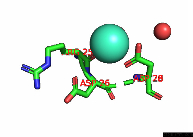
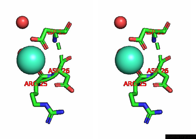
Gadolinium binding site 1 out of 3 in 7lji
Go back to
Gadolinium binding site 1 out
of 3 in the Structure of Poly(Aspartic Acid) Hydrolase PAHZ2 with Gd+3 Bound
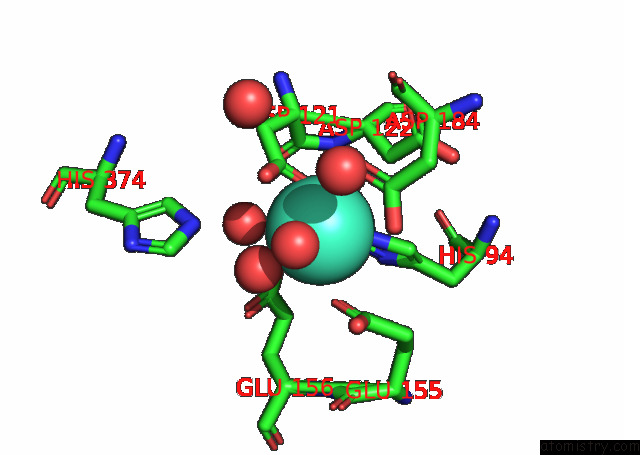
Mono view
Stereo pair view
Mono view
Stereo pair view
A full contact list of Gadolinium with other atoms in the Gd binding
site number 1 of Structure of Poly(Aspartic Acid) Hydrolase PAHZ2 with Gd+3 Bound within 5.0Å range:
|
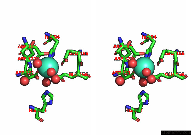
Gadolinium binding site 2 out of 3 in 7lji
Go back to
Gadolinium binding site 2 out
of 3 in the Structure of Poly(Aspartic Acid) Hydrolase PAHZ2 with Gd+3 Bound
Mono view
Stereo pair view
Mono view
Stereo pair view
A full contact list of Gadolinium with other atoms in the Gd binding
site number 2 of Structure of Poly(Aspartic Acid) Hydrolase PAHZ2 with Gd+3 Bound within 5.0Å range:
|
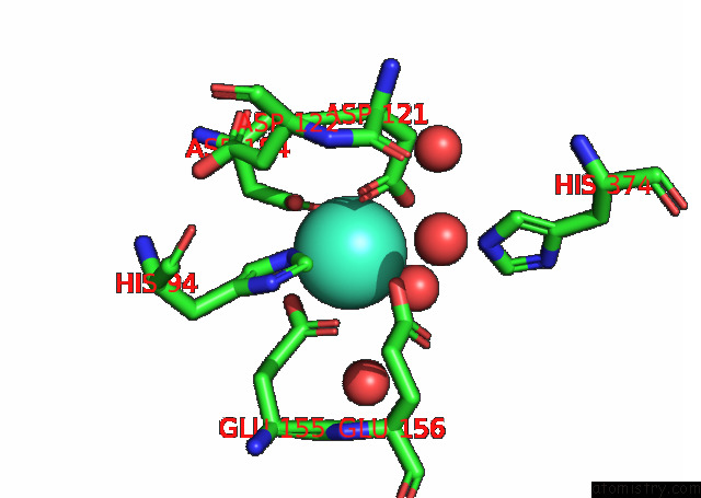
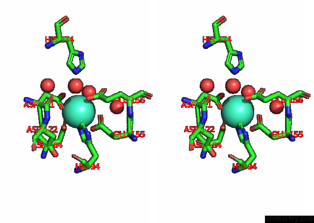
Gadolinium binding site 3 out of 3 in 7lji
Go back to
Gadolinium binding site 3 out
of 3 in the Structure of Poly(Aspartic Acid) Hydrolase PAHZ2 with Gd+3 Bound
Mono view
Stereo pair view
Mono view
Stereo pair view
A full contact list of Gadolinium with other atoms in the Gd binding
site number 3 of Structure of Poly(Aspartic Acid) Hydrolase PAHZ2 with Gd+3 Bound within 5.0Å range:
|
Reference:
C.A.Brambley,
T.J.Yared,
M.Gonzalez,
A.L.Jansch,
J.R.Wallen,
M.H.Weiland,
J.M.Miller.
Sphingomonas Sp. Kt-1 PAHZ2 Structure Reveals A Role For Conformational Dynamics in Peptide Bond Hydrolysis. J.Phys.Chem.B V. 125 5722 2021.
ISSN: ISSN 1089-5647
PubMed: 34060838
DOI: 10.1021/ACS.JPCB.1C01216
Page generated: Sat Aug 10 22:19:37 2024
ISSN: ISSN 1089-5647
PubMed: 34060838
DOI: 10.1021/ACS.JPCB.1C01216
Last articles
Zn in 9MJ5Zn in 9HNW
Zn in 9G0L
Zn in 9FNE
Zn in 9DZN
Zn in 9E0I
Zn in 9D32
Zn in 9DAK
Zn in 8ZXC
Zn in 8ZUF